The amount of bone remodeling around implants is important for a stable and predictable esthetic result. The purpose of this study was to use radiographic assessment to investigate the amount of bone remodeling that occurred after long-term functional period. A one-piece titanium device with a smooth, machined prosthetic receiving end and a rough sandblasted and acid-etched (SLA) surfaces along the entire length of the implantable screw end. The location of the smooth to rough (r/s) border is located 1 mm apical to the prosthetic margin. All devices were placed with a non-submerged surgical technique and the surgical sites were prepared with I mm deep countersink so that the device prosthetic margin is always level with the crestal bone at time of initial placement.
Vertical measurements of mesial and distal radiolucent areas adjacent to the device from digitized peri-apical radiographs were quantified from the prosthetic margin to the first bone-to-implant contact (fBIC) on the day of device placement, at insertion of prosthesis and with the most current peri-apical radiograph available for that device. 356 of these one-piece implantable devices were able to support a total of 160 fixed metal-ceramic restorations in 125 patients. 30 out of 65 single implant crowns were screw-retained the other 35 single crowns were cemented. 95 other fixed screw-retained restorations replacing all partial and edentulous conditions were supported by multiple implants.
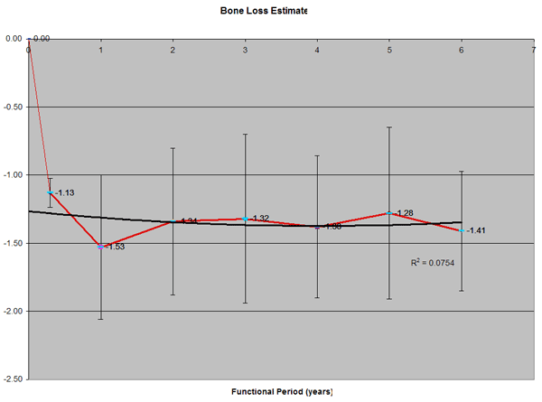
The average healing time from implant placement to insertion of prosthesis was 3.04 months, SD 1.48. A significant amount of negative bone remodeling occurred for all implants for this unload period. The initial vertical fBIC for the bone loss at time of prosthesis insertion was 1.80 mm, SD 0.79. The other time points reported are devices that have been in function for 12, 24, 36, 48, 60 and over 72 months. The results showing virtually no significant additional vertical bone losses with devices in functions for 12 months; 1.89 mm, SD 0.73; for 24 months 1.80 mm, SD 1.21; 36 months 1.89 mm, SD 1.07; 48 months 2.08 mm, SD 0.92; 60 months 1.95 mm, SD 0.92; 72 months 2.29 mm, SD 0.83); Non-paired Student t-test confirms there is no difference in vertical crestal bone loss between devices that were in functions for 12 months and 72 months at P> 0.92.
In conclusion, the bone remodeling around this one-piece device: 1) A significant amount of negative bone remodeling (bone loss) at the crestal areas occurred early (8 weeks) during the healing or unload period for all implants. 2) Crestal bone level was at a steady height to about 2 mm apical to the prosthetic margin and 1 mm apical to the r/s border. 3) The crestal bone level remains virtually unchanged up to and over 84 months. 4) There were 27.93 % of the devices in this study that appears to have positive bone remodeling (bone gain) in the crestal areas after the initial negative remodeling period.
|